Buying Group Products
Rexxam (Shin Nippon) Specular Microscope
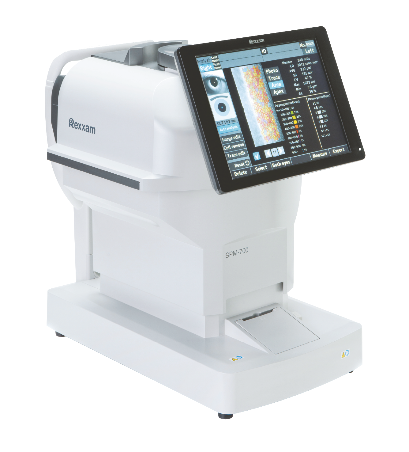
SPM-700 Specular MicroscopeDescription :
Specular microscope with easy operation and speedy analysis Easy, Speedy, and Accurate. Quick Measurements & Analysis. By simple touch panel operation, alignment is achieved automatically, images are captured continuously in 2 sec. and analyzed in 1 sec. High speed and accuracy specular microscope has been realized.
Product Specifications :
Continous Capturing of 16 Images
16 images are captured in 2 sec. with our unique zoom function and auto alignment function by touching paracenter area.
Full-auto, semi-auto and manual can be selected in the operation mode.
Speedy Analysis Function
After the measurements, the best image is selected automatically from 16 images. After selecting the best image, analysis is finished in 1 sec.
The image can be selected from 16 images manually.
Multiple Measurement Points
Total 17 measurement points including center, 6 in the paracenter and 10 in the periphery can be measured in the range of 0.25mm by 0.55mm.
Corneal Thickness Measurement
It is possible to capture the endothelial cell and to take a measurement of corneal thickness at the same time.
Edit Function
This function enables to edit the contrast, brightness and analysis result of the endothelial cell image captured. Also, it allows to remove cells, add/delete lines and divide/merge cells.
2 Manual Analyses
There are 2 manual analyses, center method and frame method.
center method
frame method
4 Types of Display Mode
The display mode can be selected from image of endothelial cell, trace display, area display and pleomorphism display.
Wide Screen
10.4 inch wide color screen.
The swivel/tilt function allows the operator to support easily the patient during operation.
Electric Chinrest
It is easy to align the eye position of the patient with the eye mark.
For More Details and Special Price for OBG Members Call 1800 - 572 -1051